

Diagnostic test
Nerve fiber skin biopsy
A nerve fiber skin biopsy is a brief office procedure that takes three tiny skin samples, about 3 millimeters wide, from the upper back, thigh and lower leg. The samples are tested for small nerve fibers and for alpha-synuclein, which makes it the gold-standard test for small fiber neuropathy and a leading method for detecting Parkinson’s disease and related conditions.
What is a nerve fiber skin biopsy?
A nerve fiber skin biopsy is a brief office procedure that takes three small skin samples. The samples are tested both for your small nerve fibers and for a misfolded protein called alpha-synuclein.
This dual analysis is the gold-standard test for small fiber neuropathy (SFN) and a leading method for detecting synucleinopathies, a group of conditions that includes Parkinson’s disease, multiple system atrophy, pure autonomic failure and Lewy body dementia.
- Tiny samples: upper back, outer thigh and outer lower leg
- 3
- Size of each sample, about a pencil eraser
- 3 mm
- Typical time until results are available
- 4–6 weeks
Why might your doctor order it?
- Burning, tingling, numbness or pins-and-needles pain
- Evaluation of diabetic neuropathy
- Neuropathic symptoms after long COVID or a concussion
- Evaluation of POTS (postural orthostatic tachycardia syndrome) or dysautonomia
- Suspected Sjögren’s syndrome or another autoimmune neuropathy
- Small fiber neuropathy with no clear cause after normal blood work
- Evaluation of Parkinson’s disease or atypical parkinsonian features
- Suspected multiple system atrophy, pure autonomic failure or Lewy body dementia
- REM sleep behavior disorder or unexplained loss of smell
- Monitoring the response to treatment over time
Some people with POTS also have small fiber neuropathy: in one study of 80 people with POTS who had skin biopsies, about 1 in 4 had a reduced number of small nerve fibers. The biopsy is often used together with autonomic testing.
What should you expect?
Before
- Continue your regular medicines unless told otherwise.
- If you take blood thinners, ask whether to pause them; most punch biopsies do not require stopping.
- Eat normally; light caffeine is fine.
- Wear loose clothing: we need access to your upper back, outer thigh and lower leg.
- Tell us if you are allergic to lidocaine or other local anesthetics.
During
Three small biopsies are taken: one from the upper back near the base of the neck, one from the outer thigh (about 4 inches above the knee) and one from the outer lower leg (about 4 inches above the ankle). Each site is cleaned and numbed with a small injection of lidocaine, and a 3 mm punch takes a tiny circular sample. The site is closed with an adhesive bandage; stitches are rarely needed.
After
Keep the bandages dry for 24 hours, then shower normally. Avoid heavy lifting and vigorous exercise for 2 to 3 days. Mild soreness is normal, and over-the-counter pain relievers help. Call us if you see signs of infection: increasing redness, swelling, discharge or fever.
What can the test show?
Nerve fiber analysis
- Epidermal nerve fiber density (ENFD): how many small nerve fibers are present
- The pattern of nerve loss across the three sites
Alpha-synuclein analysis
- Phosphorylated alpha-synuclein: protein deposits that indicate a synucleinopathy
- Helps support a diagnosis of Parkinson’s disease, multiple system atrophy or related conditions
Why three different sites?
Because the pattern matters as much as the presence.
- Three sites, three distances from the spine: upper back (proximal), thigh (middle) and ankle (distal).
- A length-dependent pattern, worse at the ankle and better at the back, is typical of diabetes and many common causes.
- A non-length-dependent pattern, similar at all three sites, suggests autoimmune, post-viral or other causes that need different treatment.
Is it safe?
A very safe minor procedure
Yes. A punch biopsy involves:
- No radiation and no IV
- Only a local anesthetic (lidocaine)
- A very low infection risk (under 1%)
- Minimal bleeding, because the samples are tiny
- Small marks that heal like a chickenpox scar
- Sites placed where clothing usually covers them
How long does it take, and where is it done?
- How long it takes
- About 5 to 10 minutes
- When results are ready
- About 4 to 6 weeks
- Where
- Walnut Creek and Vallejo
- Insurance
- Coverage depends on your plan. Please check with your insurance company.
 East Bay
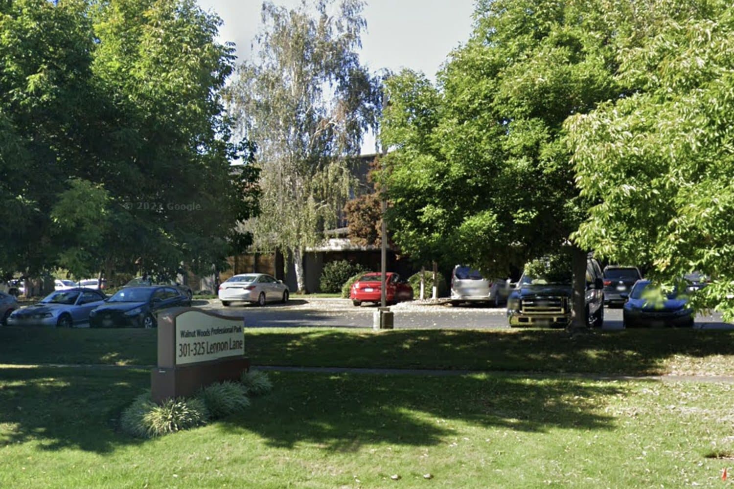
INPM Walnut Creek
301 Lennon Lane, Suite 203Walnut Creek, CA 94598
- Wed and Fri
- 8:00 AM – 5:00 PM
- Sat
- 9:00 AM – 3:00 PM
 North Bay
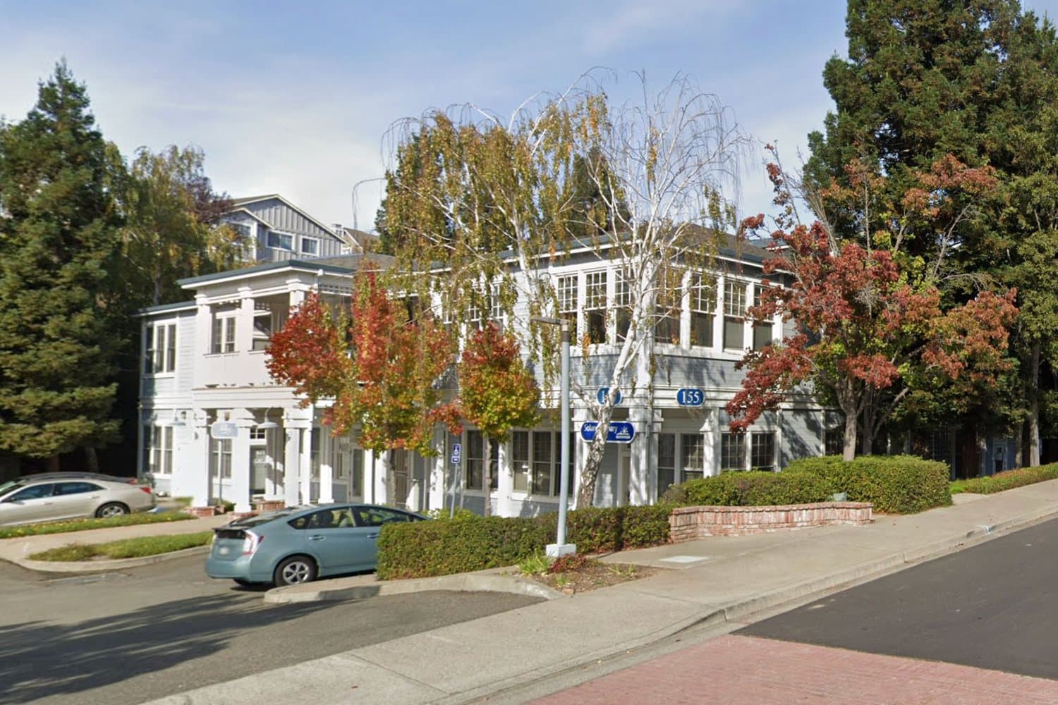
INPM Vallejo
155 Glen Cove Marina Rd, Suite 100Vallejo, CA 94591
- Mon, Tue and Thu
- 8:00 AM – 5:00 PM
Questions
Frequently asked questions
01Is a skin biopsy painful?
Each site is numbed with a small injection of lidocaine, so most people feel only that small needle. Mild soreness afterward is normal, and over-the-counter pain relievers help.
02Why are three sites sampled?
The three sites are at different distances from the spine, so the pattern of nerve loss can be compared. Loss that is worse at the ankle is typical of diabetes and many common causes, while a similar loss at all three sites suggests autoimmune, post-viral or other causes that need different treatment.
03Do I need to stop blood thinners before a skin biopsy?
Most punch biopsies do not require stopping blood thinners. Ask our office whether to pause them, and tell us about any bleeding disorder.
04When are the results ready?
Biopsy results typically take about 4 to 6 weeks to become available. Your doctor will review them with you.
Referral required
Start with a referral from your doctor
New patients are seen by referral. Your primary care physician can fax the referral to (707) 980-6692 or send it through EPIC, and our office will call you to schedule.
- Referral fax (707) 980-6692
- EPIC referral
- We call you to schedule